What Are Hippocampal Paroxysmal Discharges (HPD)?
Home » Resources / Blog / Preclinical Epilepsy - What is Hippocampal Paroxysmal Discharges (HPD)
Hippocampal Paroxysmal Discharges (HPD) are key electrophysiological events that play a central role in the study of mesio-temporal lobe epilepsy (MTLE). Understanding HPD is essential for developing and testing new antiepileptic drugs (AEDs). In this article, we explain what HPDs are, how they are recorded, and why SynapCell’s MTLE mouse model is a powerful translational tool for evaluating pharmacoresistant epilepsy.
Understanding Mesio-Temporal Lobe Epilepsy
Mesio-temporal lobe epilepsy (MTLE) represents one of the most common forms of focal epilepsies. Generally resistant to most pharmacological treatments, this type of epilepsy affects about 10% of the total epileptic population. SynapCell has an extensive expertise modeling the disease with its MTLE mouse, a translational model that mirrors human MTLE. The MTLE mouse is a non-convulsive model of focal, pharmacoresistant epilepsies. Its EEG reveals spontaneous and recurrent Hippocampal Paroxysmal Discharges (HPD), which are used as objective EEG biomarkers to reliably evaluate the effect of anti-epileptic drugs (AED) in vivo.
Breaking down the mechanisms of HPD in MTLE
Mesio-Temporal Lobe Epilepsy represents a major challenge in the clinical management of seizures. This form of epilepsy is often associated with severe cognitive and emotional impairments, and affects about 10% of the total epileptic population. In addition, MTLE becomes generally resistant to most pharmacological treatments (Engel et al., 1997).

SynapCell’s translational MTLE Mouse Mode
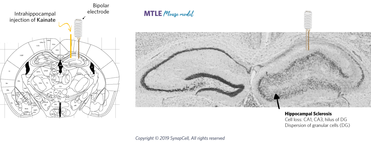
SynapCell has developed an animal model of MTLE in which, unlike most other models, dispersion of the dentate gyrus is observed (Bouilleret et al., 1999 and Riban et al., 2002). In this model, unilateral injection of a small dose of kainate (1 nmole) in the dorsal hippocampus of mice results in neuronal losses in CA1, CA3 areas and hilus as well as mossy fiber sprouting, in addition to an important dispersion and increase of granule cells of the dentate gyrus (Suzuki et al., 1995 and Mitsuya et al., 2009).
Recording and characterizing Hippocampal Paroxysmal Discharges
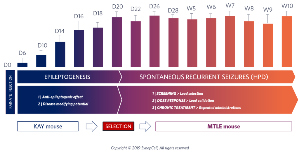
Three weeks after kainate injection, spontaneous and recurrent hippocampal paroxysmal discharges (HPD), lasting about 15-20 s, can be recorded using electroencephalography (EEG), concomitantly with behavioral arrest and/or automatisms.
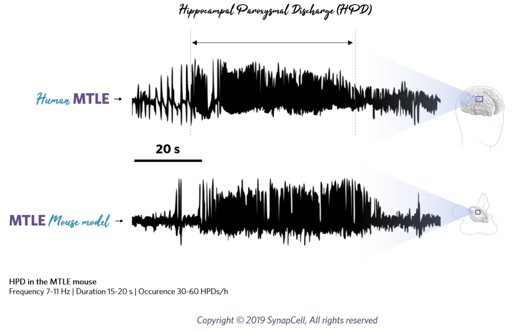
These focal seizures remain stable during the rest of the animal life and occur regularly (about 40/h) when the animals are in a state of quiet wakefulness (Riban et al., 2002 and Arabadzisz et al., 2005). During the first two weeks following kainate injection, before the occurrence of HPD, interictal spikes progressively occur by bursts of increasing duration
Evaluating antiepileptic drug efficacy using HPD
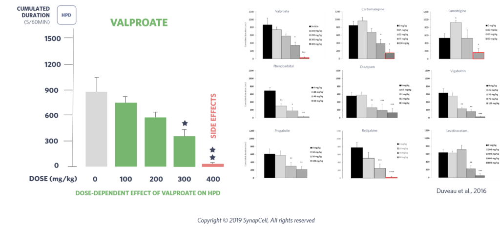
More importantly, this model is resistant to most classical antiepileptic drugs: valproate, carbamazepine and lamotrigine can suppress the hippocampal discharges upon acute treatment only at high doses and with side effects (Riban et al., 2002; Duveau et al., 2016). We have shown that HPD are dose-dependently suppressed by acute administration of diazepam. However, recently developed AEDs, such as pregabalin, levetiracetam or vigabatrin were shown to be active on this model with no side effects (Duveau et al., 2016).
Why HPD is a critical Biomarker for translational epilepsy research ?
Altogether the histological, electrophysiological, behavioral and pharmacological features, in addition to a species-appropriate latent period following the initial insult, validate this model of pharmaco-resistant MTLE. Indeed, it fulfils most criteria recently proposed for an efficient preclinical development of antiepileptic and prophylactic therapeutic strategies for MTLE (White, 2003).
FAQ - Hippocampal Paroxysmal Discharges
What are Hippocampal Paroxysmal Discharges used for in preclinical research?
They serve as objective electrophysiological biomarkers to quantify seizure activity and evaluate antiepileptic drug responses in vivo.
How are HPDs detected in MTLE models?
HPDs are recorded using intracranial EEG in freely moving animals, typically four weeks after kainate-induced hippocampal injury.
Why is the MTLE mouse model relevant for pharmacoresistant epilepsy?
Because it exhibits spontaneous, stable HPD and mirrors key histological and electrophysiological features of human MTLE.
Do conventional antiepileptic drugs suppress HPD?
Only partially and generally at high doses, leading to adverse side effects. This highlights the pharmacoresistant profile of the model.
What makes HPD a translational biomarker?
Its stability, reproducibility, and cross-species relevance allow the assessment of drug effects using endpoints that reflect human MTLE mechanisms.